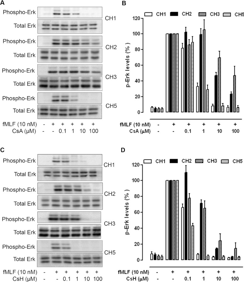
Figure 4. Inhibition of ERK1/2 phosphorylation by CsA and CsH.
CHO-Gα16-FPR1 cells were treated with either CsA (A and B) or CsH (C and D) for 15 min as indicated and stimulated with fMLF (10 nM) for 5 min. Cell lysates were prepared, separated by SDS/PAGE and blotted with specific anti-phospho-ERK1/2 antibodies as described in the Experimental section. The total protein kinase levels in cell lysates were determined by blotting with antibodies against the non-phosphorylated ERK1/2, as shown at the bottom of each panel. Images displayed in (A) and (C) are representative of at least three independent experiments with similar results. The results in (B) and (D) are the means±S.E.M. of three independent experiments, with the ERK1/2 phosphorylation induced by fMLF set as 100%.

