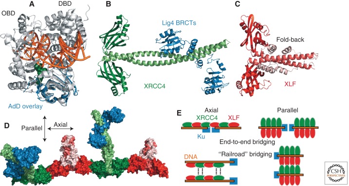
Figure 2.
DNA ligase IV assembly. (A) The adenylation domain (AdD) of Lig4 (blue, PDB 3VNN) (Ochi et al. 2012) is superimposed on a structural representation of Lig1 bound to a DNA nick (light gray, PDB 1X9N) (Pascal et al. 2004) as a surrogate model of how Lig4 might bind DNA. (OBD) Oligonucleotide/oligosaccharide binding domain; (green) 5′ AMP; (orange) DNA. (B) The human XRCC4 homodimer bound to the Lig4 tandem BRCT repeat region (PDB 3II6) (Wu et al. 2009). (C) Human XLF homodimer (PDB 2QM4) (Li et al. 2008b). (D) Surface representation of the XRCC4–XLF axial filament with a bound Lig4 BRCT region, created by superimposing PDB 3II6 onto PDB 3RWR (Andres and Junop 2011). (Blue) Lig4; (shades of green) XRCC4; (shades of red) XLF. (E) Idealized models of DNA engagement and end bridging by XRCC4–XLF multimers, colored the same as in D. “Axial” and “parallel” refer to the orientation of XRCC4–XLF interactions that drive the assembly.

