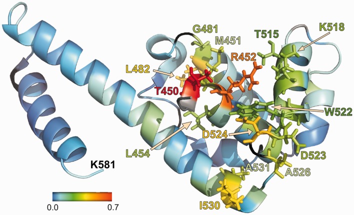
Figure 6.
NMR analysis identifies amino acids of DnaG-C involved in binding to the C-terminus of SSB. Changes in chemical shift for the amino proton and nitrogen resonances in the backbone of DnaG-C after addition of SSB-Carb peptide. The shift of the resonances was calculated using the formula shift = sqrt{[Δδ(1H)]2 + [Δδ(15N)/10]2} (where Δδ is the difference in chemical shift in the respective dimensions), and is coloured according to the scheme given in the figure. Amino acids with a shift of >0.2 ppm are shown with side chains using the same colour code. Proline residues and unassigned amino acids are shown in black. Where no shift could be determined (because of peaks missing in the spectrum without peptide, see text), the amino acid is shown in grey. Amino acids 435–446 are missing in the pdb-file 2HAJ (33) and are thus not shown here. Of these residues, however, only L446 did show a significant shift (0.21 ppm).

