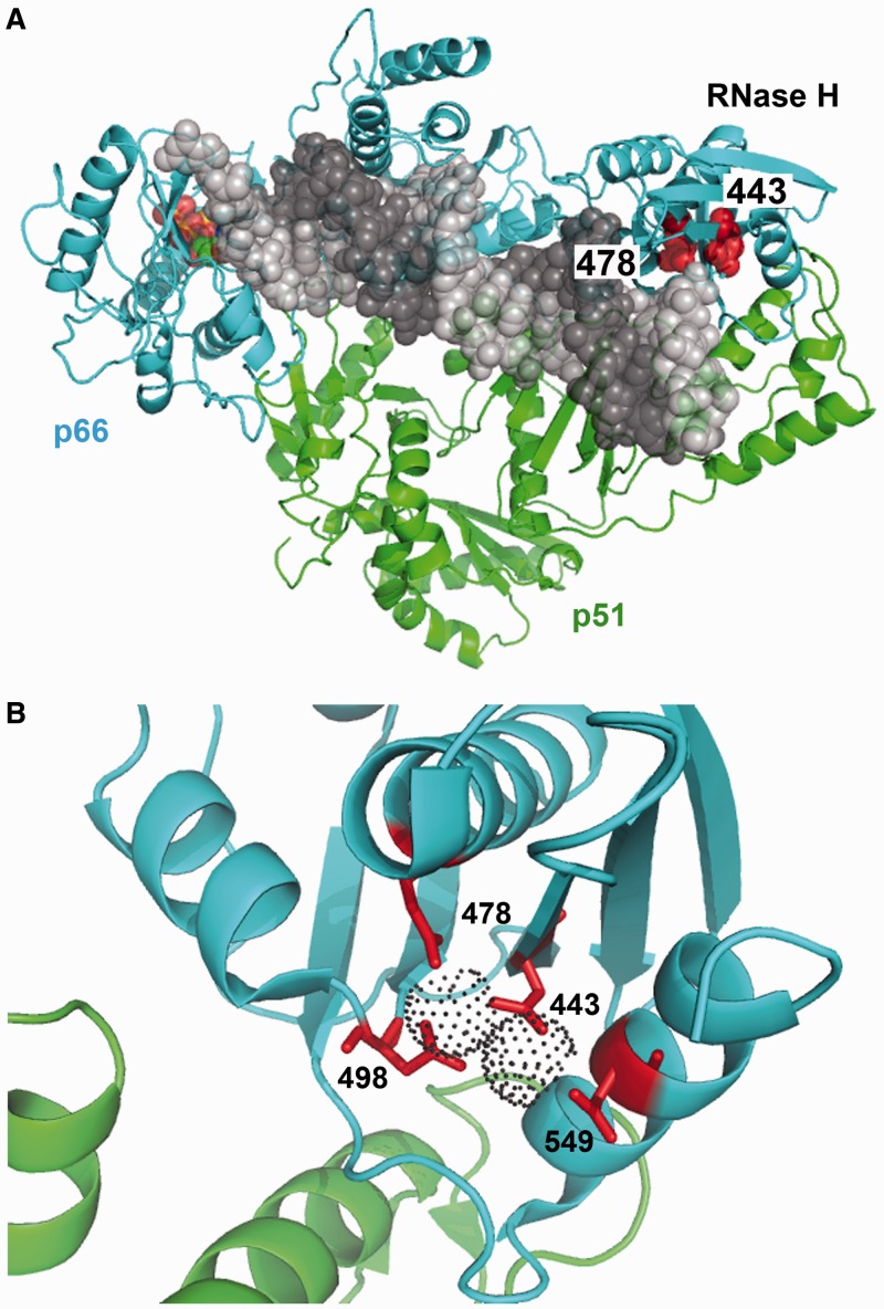
Figure 4.
Crystal structure of HIV-1 RT showing the location of RNase H active site residues. (A) Ternary complex of HIV-1 group M subtype B RT, double-stranded DNA and dTTP [PDB coordinates from Huang et al. (39), PDB code 1RTD]. The RT subunits are represented by cyan and green ribbons. The template and primer strands are shown in light and dark grey sphere models. The incoming dNTP is shown in orange, and the side-chains at positions 443 and 478 are shown in red. (B) Close-up view of the RNase H active site showing the location of Asp443, Glu478, Asp498 and Asp549 and the coordinating metal ions (dot surfaces) [PDB coordinates from Su et al. (40), PDB code 3LP1].

