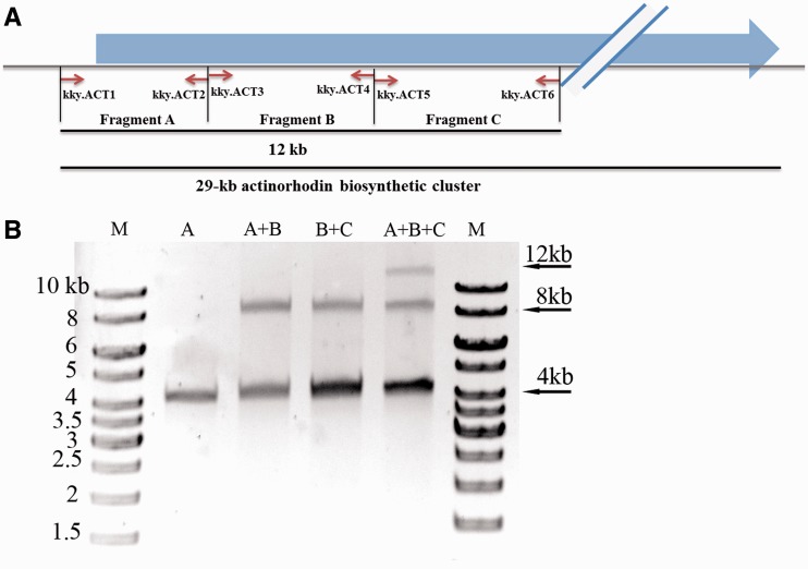
Figure 2.
In vitro seamless assembly of 3 PCR amplicons. (A) The schematic diagram of the PCR amplicons. The fragments A (4092 bp), B (4000 bp) and C (4084 bp) were amplified from the actinorhodin biosynthetic cluster with designed primers (Supplementary Table S1). (B) The PCR amplicons were digested with MspJI and used for ligation at 16°C for 2 h. Fragments A, B and C were indicated on each lane. ‘M’ stood for 1-kb DNA ladder, and the sizes of the ladder were labelled.

