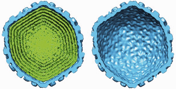
Figure 1.
Cutaway views of the wt lambda phage cryo-EM reconstructions on complete DNA packaging (left) and after ejection of the DNA (right). The size and structure of the capsid shell (blue) remains unchanged after DNA ejection. Spacing between the outermost layers of the DNA (green) can be observed in the fully packaged phage reconstruction, and the DNA becomes more disordered closer to the centre of the capsid. The d-spacings between the DNA layers inside the capsid were determined by computing 3D cryo-EM reconstructions of the phage particles (see ‘Materials and Methods’ section). Owing to the icosahedral symmetry imposed during the reconstruction, concentrically packed DNA within the capsid becomes shells of density. The central slice of each reconstruction was extracted along the 5-fold symmetric axis, providing a cross-section of density in which the capsid and packaged genome appear most circular.

