Figure 4.

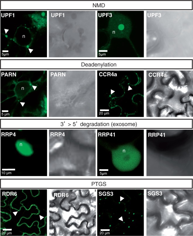
Subcellular localization of NMD, deadenylation, exosome and PTGS components. Confocal sections and their corresponding bright-field images of N. benthamiana leaves expressing the indicated proteins fused to GFP. The arrowheads indicate cytoplasmic foci, whereas ‘n’ labels the nucleus. Scale bars are shown on the images.
