Figure 3.
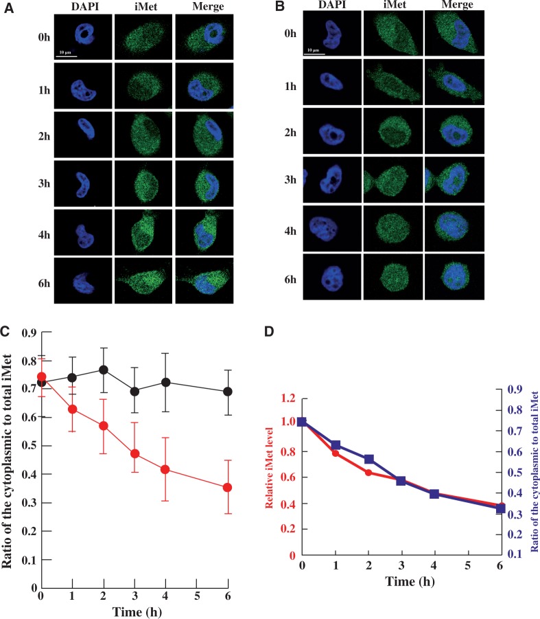
Heat stress caused nuclear accumulation of tRNA(iMet). Nuclear accumulation of tRNA(iMet) was monitored by FISH in HeLa cells at 37°C (A) and 43°C (B) for the time indicated. (C) Ratio of cytoplasmic to nuclear tRNA(iMet) at 37°C (black) and at 43°C (red) is shown. Quantification of fluorescence was performed in 40 cells per hour by using the LAS program (Leica) and the results were averaged. Data represent the mean±SD (n = 40). (D) Superposition of degradation and accumulation of tRNA(iMet). The circles show the degradation of tRNA(iMet). The squares show accumulation of tRNA(iMet) from the cytoplasm to the nucleus.

