
Figure 7.
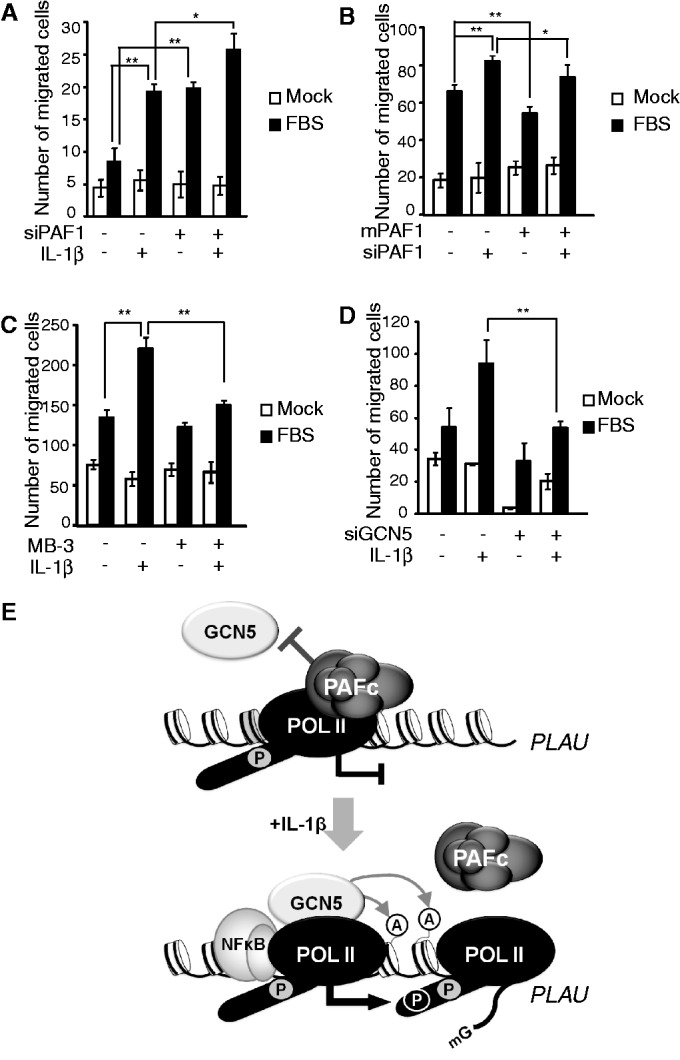
The counteraction between PAF1 and GCN5 controls the IL-1β–induced migration of HepG2 cells. (A–D) HepG2 cells with or without IL-1β stimulation were loaded onto the upper chamber, and cell migration towards the FBS-containing lower chamber during 24 h was determined using Diff-Quick staining. Three independent wells of a Boyden chamber were used to count the migrating cells. (A) Cells transfected with control or PAF1 siRNA were analyzed using a Boyden chamber assay. (B) Cells were co-transfected with control or PAF1 siRNA and empty vector or mPAF1 expression vector, and cell migration was determined without IL-1β stimulation. (C) HepG2 cells were pre-treated with MB-3 for 24 h, and cell migration was determined with or without IL-1β stimulation. (D) Cells transfected with control or GCN5 siRNA were analyzed using a Boyden chamber assay. (A–D) Mean ± SD of three experiments was presented. (E) A proposed model of PAF1-mediated gene repression. Without stimulation, chromatin-bound PAFc blocks the recruitment of GCN5 and represses RNA Pol II paused gene expression. Upon IL-1β stimulation, the GCN5-dependent acetylation of histones releases PAFc from chromatin and facilitates continued productive transcription.