Figure 1.
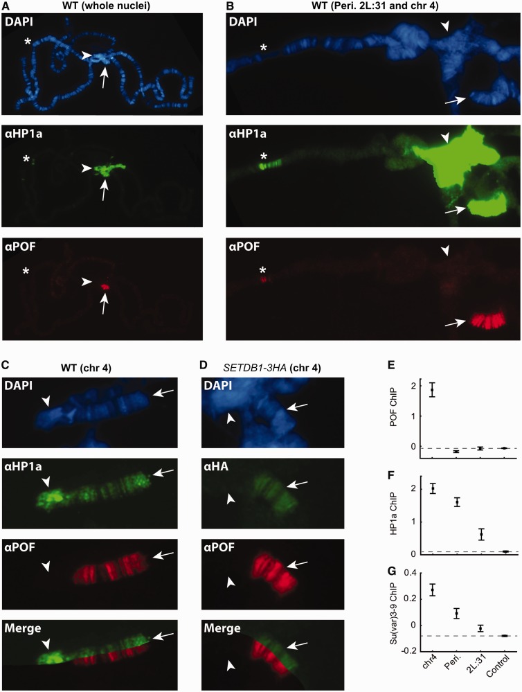
HP1a, SETDB1 and POF binding overlaps on chromosome 4, occasionally POF overlaps with HP1a binding on region 2L:31. (A) HP1a (green) and POF (red) localization on a whole wild-type polytene chromosome. (B) Close-up image of pericentromeric, chromosome 4 and 2L:31 regions. The arrow indicates chromosome 4, arrow head indicates pericentromeric region and asterix indicates cytological region 2L:31. (C) POF and HP1a binding on chromosome 4. (D) POF and HA (for detection of HA-tagged SETDB1) staining in red and green, respectively, on chromosome 4 in a SETDB1-3HA third-instar larva. The arrow indicates chromosome 4 and arrow head indicates pericentromeric region. DNA is stained with DAPI (blue). (E–G) Mean exon binding value (log2 scale) of POF (E), HP1a (F) and Su(var)3-9 (G) for all active genes within chromosome 4, pericentromeric regions, 2L:31 region and control region (whole chromosome 3R) (n =50, 68, 56 and 1753, respectively). Dashed lines represent binding levels in the control region (chromosome 3R) and error bars indicates the 95% confidence interval.

