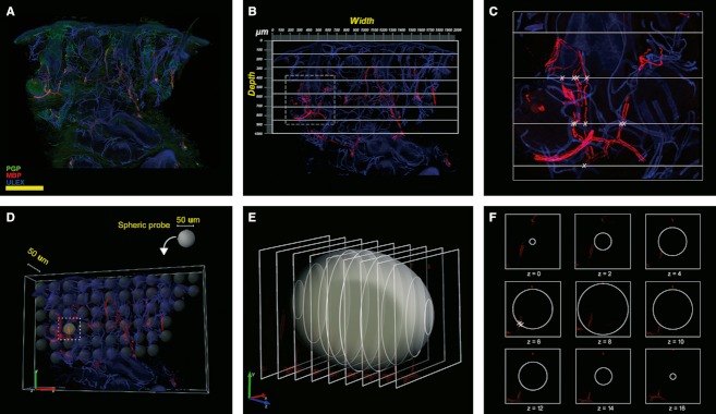
Fig. 2.

Images exemplifying the methods used to quantify myelinated fibers. In green, PGP; in red, MBP; in blue, Ulex Europaeus agglutinin A. (A–C) Method used to quantify MF. (D–F), the unbiased stereological method used as gold standard in the field. (A) MBP/PGP/Ulex triple-stained 5× non-confocal image. Myelinated fibers are in red, axons are in green and epidermis and endothelium are in blue. (B) Application of a calibrated grid (2 × 1 mm) with a series of seven equidistant parallel lines on the same image using only the red and blue channels. (C) The area of tissue included in the square in (B) at higher magnification. The intersections between grid lines and fibers are marked with an X. Scale bar: 400 μm. (D) Distribution of virtual ‘space ball’ probes within the tissue. (E) Intersections between the probe and the confocal optical sections. (F) Intersections between nerve fibers and the probe are marked with an X.
