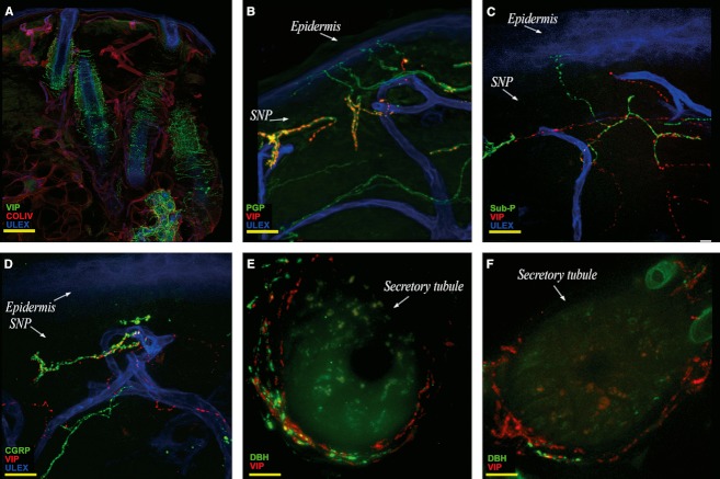
Fig. 5.
Confocal images showing cutaneous autonomic innervation. (A) Collage showing VIP fiber distribution in the dermis around dermal annexes. (B,C,D) VIP-ir fibers in the SNP, in close contact with Sub P-ir fibers (C) and CGRP-ir fibers (D). Facial noradrenergic sudomotor innervation is consistently well represented (E) compared with non-facial body sites (F). Images are from the third (B–D) and the second trigeminal branch (A–E) and from the thigh (F). In green, VIP (A), PGP (B), Sub P (C), CGRP (D), DβH (E,F); in red, collagen IV (A), VIP (B–F); in blue, Ulex Europaeus agglutinin A. Scale bar: 160 μm (A), 100 μm (B); 50 μm (C,D); 20 μm (E,F).

