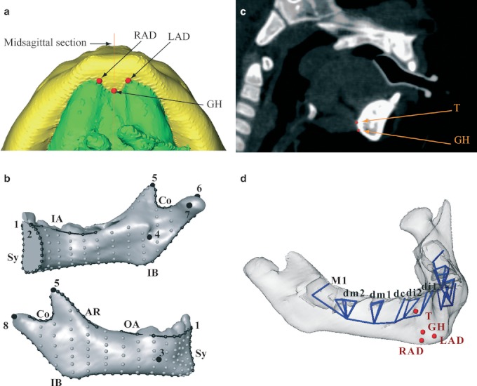
Fig. 3.

(a) Reconstruction of the mandibular corpus and the suprahyoid muscles (inferior view) together with the landmarks RAD (anterior belly of the right digastric muscle), LAD (anterior belly of the left digastric muscle) and GH (genio-hyoid muscle). (b) Landmark template on the right hemimandible of a 1-year-old specimen. Anatomical landmarks are large black dots, curve semi-landmarks are smaller black dots connected by black lines, and surface semi-landmarks are gray dots. Names of the landmarks and curves are as in Table 1. (c) The landmarks T (tongue) and GH on a sagittal section of a patient. (d) The mandibular template was supplemented with 38 deciduous tooth landmarks (blue vertices) and four muscle landmarks (red dots).
