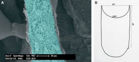
Fig. 2.

(A) Vascular canal cast from a mono-layer infiltration depth of the lacunar-canalicular network. The light-blue tinted area corresponds to a segment of the hemi-canal surface used to estimate the density of the lacunar cast first layer (n mm−2). (B) Diagram illustrating the method of assessment of the hemi-canal segment surface (nr.h). It was assumed that the transverse section of the canal was a regular circumference.
