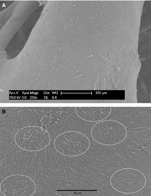
Fig. 4.

(A) Cast of a bare vascular canal where the resin penetrated inside the first tract of the canalicula openings into the canal lumen. The collagen fibril pattern is reproduced by the cast showing the ordered, parallel disposition with different angles with the canal axis. Scattered lacunae have been infiltrated by the progression of the resin within the lacunar-canalicular system. (B) Detail of (A) showing the initial cast of the canalicula connecting the canal lumen with the intra-osteonal system. The oval areas where they are more densely packed correspond to the positions of the osteocyte not yet infiltrated by the resin.
