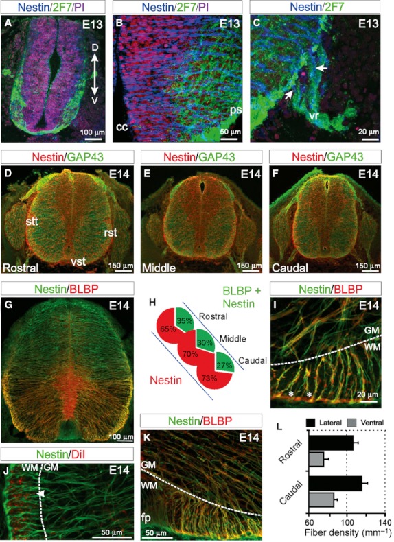
Fig. 2.

The organisation of radial neuroepithelial and radial glial cells at the onset of white matter (WM) formation in the spinal cord. (A) At E13, nestin-immunoreactive neuroepithelial cells are present ventrally and dorsally. 2F7-immunoreactive mature and immature neurons and axons are present in the presumptive ventral horn and WM, respectively, in the presumptive lateral WM and crossing over the ventral midline. (B) Nestin-immunoreactive radial cells extend from the central canal to the pial surface through the emerging ventral grey matter (GM) and WM. (C) In the ventrolateral spinal cord, some nestin-immunoreactive processes extend past the pial surface and appear to enter the ventral root (vr; arrows), possibly guiding motor axons exiting the spinal cord. (D–F) At E14, the emerging ventral and lateral WM tracts are present in the rostral, middle and caudal spinal cord. These tracts correspond to the growing vestibulospinal tracts (vst) ventrally, the spinothalamic tracts (stt) and the reticulospinal tracts (rtt) laterally. A band of axons entering from the dorsal root are evident dorsally. Nestin-immunoreactive processes radiate from the central canal to the pial surface in the rostral, middle and caudal spinal cord. (G) At E14, brain lipid-binding protein (BLBP) is coexpressed with nestin in the ventral portion of the spinal cord in radial glial processes. (H) The proportion of BLBP and nestin expressing radial glial cells compared with nestin expressing radial neuroepithelial cells decreases along a rostral to caudal gradient. (I) Two-hundred-micrometer-thick slices show how nestin- and BLBP-immunoreactive radial glial cells ventrally adopt a uniform orientation as they pass through the GM/WM interface (dotted line) and radiate toward the pial surface. Occasionally, spaces are observed between radial glial cell processes that persist through the thickness of the slice (asterisks). (I–K) Radial glial cells often branch at the GM/WM interface (dotted lines) resulting in an increased number of processes entering the presumptive WM. DiI labelling of axons shows how discrete fascicles seem to be contained within channels of nestin-immunoreactive processes in the WM (arrowhead in J). (K) Near the ventral floor plate (fp), nestin- and BLBP-immunoreactive radial glial cell processes display a striking change of direction as the transition from the GM/WM interface (dotted line) to the pial surface. (L) The densities of radial glial cell processes in the dorsal and lateral WM are maintained throughout the rostrocaudal axis of the spinal cord. (Images A–F are confocal projections of 15–20 images captured at 0.5-μm intervals, images G, I–K are confocal projections of 400 images captured at 0.5-μm intervals. Scale bars are in microns as indicated. Dorsoventral orientations are consistent for all images as indicated in A).
