Fig. 3.

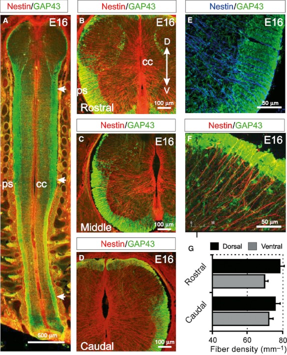
Radial glial cells coursing through the WM display a high degree of organisation in all regions of the E16 spinal cord. (A) Longitudinal section of an E16 embryo showing how GAP43-immunoreactive axon tracts are present the entire length of the spinal cord (arrows). Nestin-immunoreactive radial glial cells radiate from the central canal to the pial surface at all levels. (B–D) At E16, GAP43 expression is increasing in the ventral and lateral WM of the rostral, middle and caudal spinal cord. The presumptive WM of the dorsal horn and the dorsal funiculi is also emerging at all levels of the spinal cord. Nestin-immunoreactive radial glial cells radiate from the central canal to the pial surface both dorsally and ventrally. (E,F) Radial glial processes appear evenly distributed in the ventral (E) and dorsal (F) WM. (G) The densities of radial glial processes in the ventral and dorsal WM are maintained throughout the rostrocaudal axis of the spinal cord. (All images are confocal projections of 15–20 images captured at 0.5-μm intervals. Scale bars are in microns as indicated. Dorsoventral orientations are consistent for images B–F as indicated in B).
