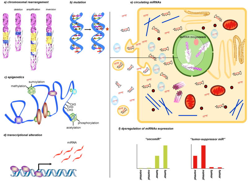
Figure 1. miRNA dysregulation in cancer.
Dysregulation of miRs expression in cancer compared to the normal tissues of origin is a general phenomenon that has been largely characterized in almost all neoplasia. Global repression of miRNAs expression in cancer cells is believed to induce an undifferentiated phenotype. Indeed the increase of specific miRs, the “oncomiRs”, confers aggressiveness and resistance to cell death. In the figure, we depicted the main processes involved in miRNA dysregulation, such as a) chromosomal alterations of the miRNA genes, b) DNA point mutations, c) epigenetic mechanisms or d) alterations in the machinery responsible for miRNA production. e) In addition to their intracellular functions, recent studies have demonstrated that miRNAs can be released or leaked from cancer cells and circulated in a remarkably stable form within blood. Many studies have demonstrated the circulating miRNA levels correlate significantly with cancer progression, therapeutic response, and patient survival. As RISC-associated, microvesicles-related or as a free miRNAs or pre-miRNAs, the function of circulatory miRNAs is largely unknown. Many questions are still missing an answer: are circulating miRNAs a result of leakage from cancer cells or active release? Besides bio-markers, can miRNA drive or activate molecules that help the body defend against cancer? f) Representation of the miRNA modulation in the neoplastic tissues compared to normal tissues of origin: an “oncomiR” upmodulation and a “tumor-suppressor miR” repression are shown in the two graphics.

