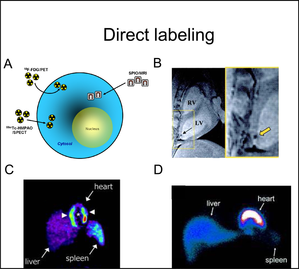
Figure 1. Direct cell labeling strategies.
A, labeling agents (for either magnetic resonance or radionuclide imaging) are first introduced into the stem cells exogenously, and are then transplanted to the tissue and/or organ of interest. Non-invasive imaging is subsequently performed. B, 2.8×10⁷ MSCs, labeled with super paramagnetic particles (Feridex, 25 µg Fe/mL), were imaged, after direct transmyocardial delivery, using a 1.5T MRI. The black signal (yellow arrow) represents the super paramagnetic signal, which has been used to monitor the delivery of stem cells. C, 1.25×10⁸ BMCs, labeled with 18F-FDG (100MBq), were delivered to the myocardium via intracoronary injection, and then imaged using PET. The white arrowheads point to the transplanted cells in the heart. There is also liver and spleen uptake (route of tracer elimination). D, 8×10⁸ BMCs were labeled with 99Tc-HMPAO (100MBq/1×108 cells) and infused via intracoronary injection to patients with chronic ischemic cardiomyopathy and imaged with SPECT at different times after delivery (shown is a representative image obtained one hour after cell delivery).
Abbreviations: SPIO: superparamagnetic iron oxide particles, 18F-FDG: 18F-fluorodeoxyglucose, 99Tc-HMPAO: Tc99m-hexamethylpropylenamineoxime, MSCs: mesenchymal stem cells, MRI: magnetic resonance imaging, PET: positron emission tomography, SPECT: single-photon emission computed tomography, RV: right ventricle, LV: left ventricle. Adapted from Kraitchman et al. Circulation 2003 13;107(18):2290–3, Gousettis et al. Stem Cells 2006; 24: 2279–2283, and Hofmann et al. Circulation 2005 111: 2198–2202 with permission.

