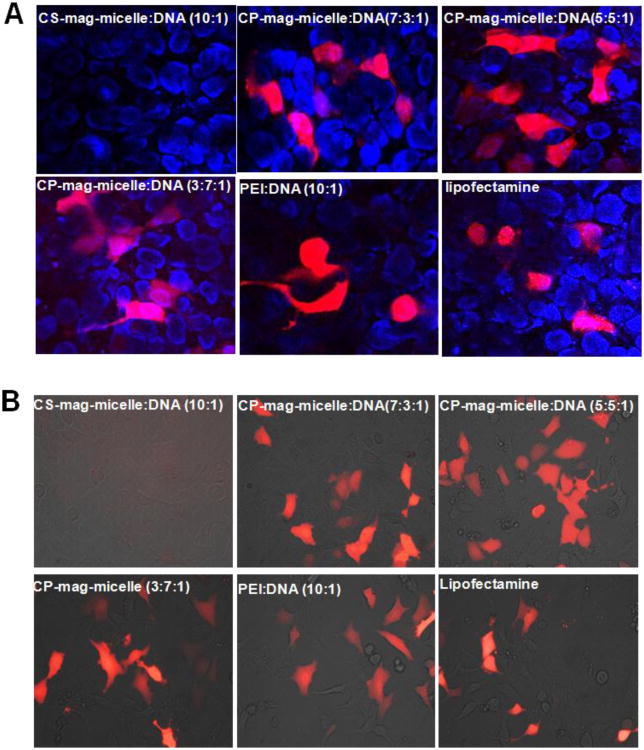
Figure 5.
Cells were transfected with indicated NPs complexed with Tomato red-fluorescent protein plasmid. Forty-eight hours after transfection, tomato protein expression was examined. Confocal microscopic images (200×) of HEK293. Nuclei were stained with DAPI (A). Fluorescence images (400×) of 3T3 cells (B).

