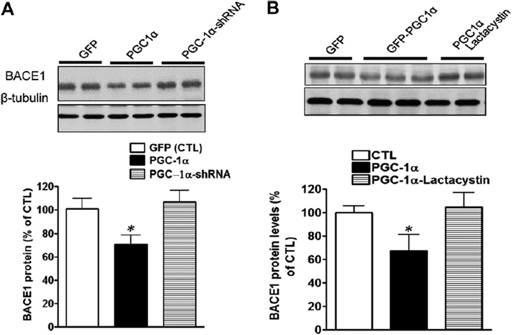
Fig. 5.
PGC-1α expression promotes BACE1 degradation. (A) Primary hippocampal-cortical neurons derived from Tg2576 embryos at 14 days in vitro were infected by adenoviral-GFP PGC-1α, scramble PGC-1α-shRNA, or PGC-1α-shRNA, respectively, 72 hours after infection, cell lysates were collected and analyzed via Western blot using anti-BACE1 antibodies. BACE1 levels were quantified and normalized against the level of β-tubulin and plotted as percentage of CTL. Data are expressed as mean±standard error of the mean (n=5). * p<0.05 compared with CTL group. Inset represents PGC-1α immunoreactive signals. (B) The degradation of BACE1 caused by PGC-1α expression on was blocked by lactacystin treatment (5 µM). Data are expressed as mean ± standard error of the mean (n = 5). * p < 0.05 compared with CTL group. Inset represents BACE1 immunoreactive signals. Abbreviations: BACE1, β-secretase; CTL, control; GFP, green fluorescent protein; PGC-1α, peroxisome proliferator-activated receptor-γ coactivator 1α; shRNA, small hairpin RNA.

