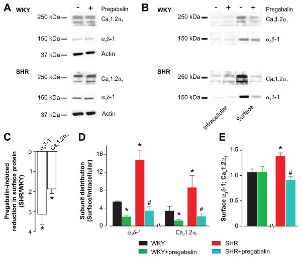
Figure 3.
Pregabalin reduces surface expression of α2δ-1 and CaV1.2α1 channel proteins more effectively in arteries of hypertensive rats than in controls. A, Representative Western blot illustrating that pregabalin does not change total (whole arterial) α2δ-1 and CaV1.2α1 proteins in WKY and SHR arteries. B, Representative Western blots illustrating pregabalin (24 h)-induced changes in surface and intracellular α2δ-1 and CaV1.2α1 proteins. Blots were cut at 75 kDa to allow probing for α2δ-1 and CaV1.2α1. C, Pregabalin reduced surface α2δ-1 and CaV1.2α1 proteins more in SHR than WKY arteries. D, Mean data illustrating α2δ-1 and CaV1.2α1 subunit distribution in WKY and SHR arteries and regulation by pregabalin. E, Surface α2δ-1 to CaV1.2α1 and modulation by pregabalin. Pregabalin concentration in all figures was 100 μmol/L. * indicates P<0.05 compared with untreated WKY and # indicates P<0.05 versus untreated SHR rat arteries (n=4–5 each for untreated and pregabalin-treated WKY and SHR).

