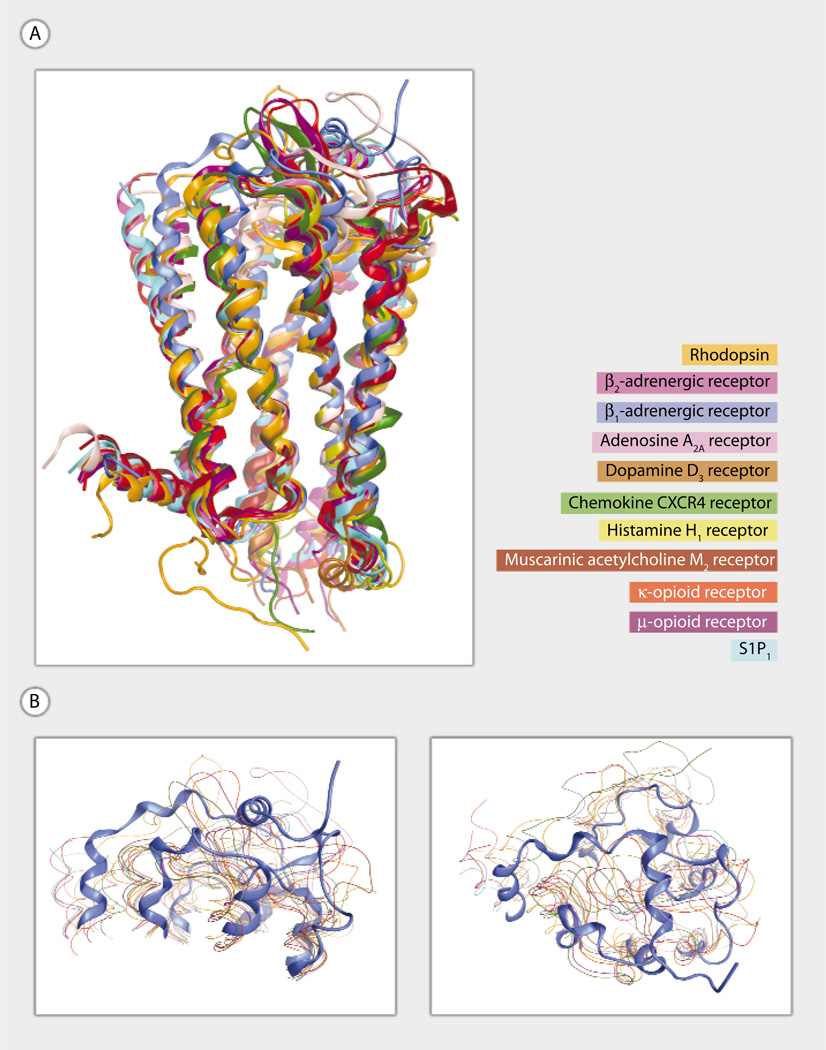
Fig. 1.
(A and B) Superposition of 11 crystallized GPCR family members. Rhodopsin (PDB ID 1F88) (16), β2-adrenoceptor (PDB ID 2RH1) (12), β1-adrenoceptor (PDB ID 2VT4) (17), adenosine A2A (PDB ID 3EML) (18), dopamine D3 (PDB ID 3PBL) (19), chemokine CXCR4 (PDB ID 3OE0) (20), histamine H1 (PDB ID 3RZE) (21), muscarinic acetylcholine M2 (PDB ID 3UON) (22), κ-opioid (PDB ID 4DJH) (23), and μ-opioid (PDB ID 4DKL) (24) are shown using ribbon (A) and line (B) representations. S1P1 (PDB ID 3V2Y) (3) is shown using a ribbon representation in all panels. The muscarinic acetylcholine M3 (PDB ID 4DAJ) (25) structure is not shown. Ribbon representations are oriented with extracellular segments at the top of the figure and transmembrane domain 1 on the left (A). T4 lysozyme replacements for IL3 are not shown. (B) Two views of the extracellular segments are shown to emphasize the unique extracellular architecture of the S1P1 receptor.

