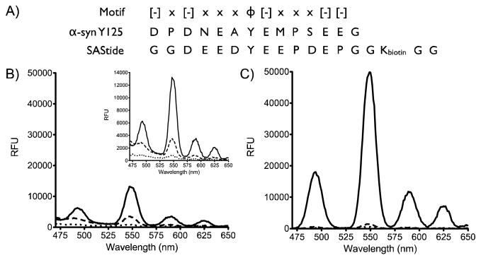
Figure 1.

Detection of phosphorylation of SAStide through terbium luminescence. A) Sequence alignment of a peptide from α-synuclein, an atypical terbium sensitizing peptide, and SAStide. B) Steady-state luminescence and C) time-resolved luminescence emission plots for SAStide and pSAStide. pSAStide (—) SAStide (---) and Tb (· · ·). Spectra were collected from 15 μM peptide in the presence of 100 μM Tb3+ in 10mM HEPES, 100mM NaCl, pH 7.0, λex=266nm, 1000ms collection time, 50 μsec delay time and sensitivity 180. Data represent the average of experiments performed in triplicate.
