
Figure 3. In vitro expression of Slit molecules impairs neural crest cell migration.

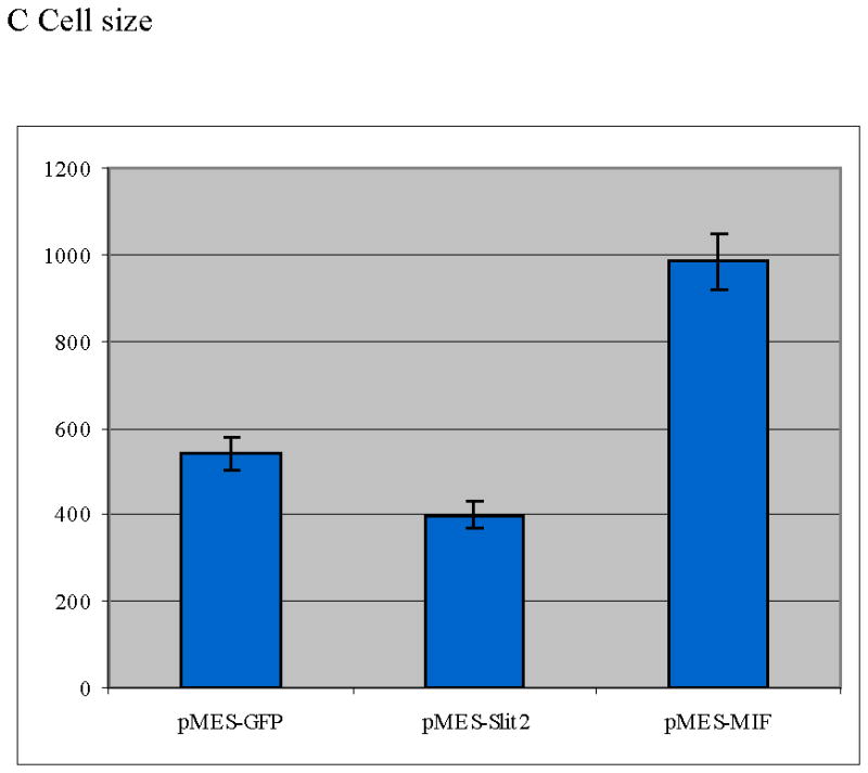
Neural crest cells were cultured in vitro and chemically transfected with GFP. A Bar graph scoring cell shape of neural crest cells after electroporation (T-test: p<0.005, N=680 per each treatment). There were far fewer neural crest cells showing a migratory/mesenchymal morphology compared with GFP cells. B Morphology of neural crest cells chemically transfected with GFP, Slit2-GFP or MIF-GFP. C Graph showing individual cell area (y axis corresponds to μm2 of cell surface) for neural crest cells after transfection. Cells expressing Slit2-GFP were significantly smaller than control-GFP or MIF-GFP expressing cells (T-test: p<0.0004 T-test, N=60 cells per each treatment).