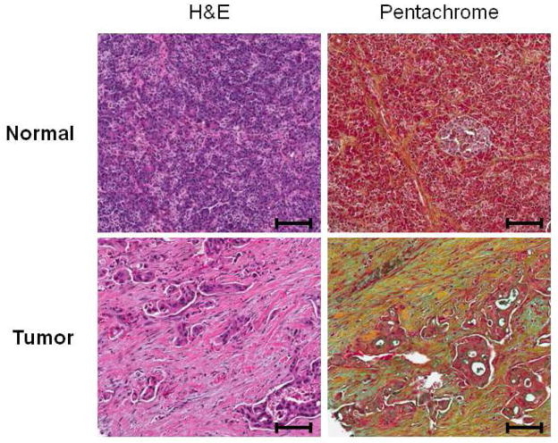
FIGURE 2.
Histochemical analysis of human pancreatic tissue samples. Both normal and tumor pancreatic tissues were subjected to hematoxylin/eosin (H&E) and pentachrome (Russell-Movat’s) staining analysis. H&E analysis reveals increased proliferation of the pancreatic stellate cell population in the tumor tissues relative to normal tissues. Pentachrome analysis also demonstrates increased collagen expression in the stromal compartment in tumor tissue relative to normal tissue. Pentachrome staining: Green/blue=mucins, Yellow=collagen, Red=muscle/fibrinoid. Scale bar=100 μm

