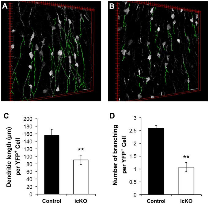
Figure 6. ERK5 deletion inhibits neuronal maturation in the OB.
(A–B) Representative 3-D images of YFP+ cells in the OB. The OB sections were cut at 20 µm. Dendrites were highlighted by green color using Simple Neurite Tracer. Images were created by the 3D viewer of ImageJ. Scale bars represent 25 µm. (C) Average dendritic length of YFP+ cells in the OB was measured. (D) Quantification of the average number of dendritic branching of YFP+ cells. n = 3 individual mouse brains and olfactory bulbs per group. **, p<0.01.

