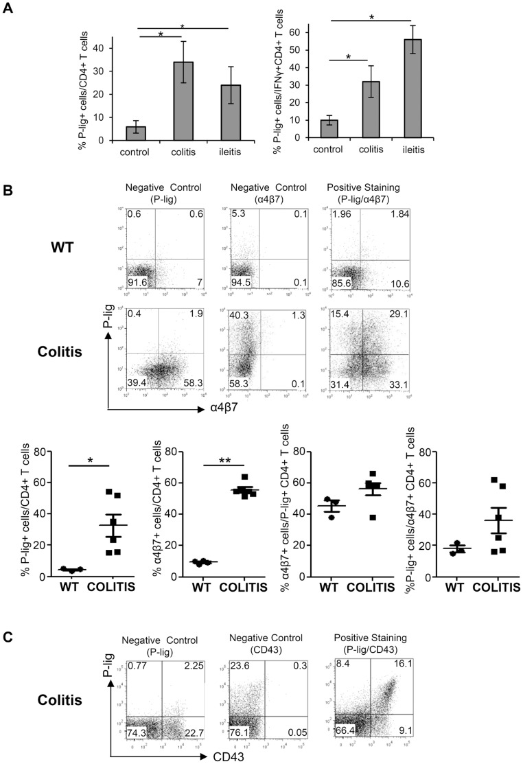
Figure 1. Coexpression of P-lig and α4β7 on a major subset of CD4+ T cells in MLN during intestinal inflammation.
A) The percentage of P-lig+ cells/CD4+ T cells (left panel) and the percentage of P-lig+ cells/IFNγ+CD4+ T cells (right panel) in MLN of healthy mice (control), SCID mice after induction of colitis (colitis) and mice orally infected with 10 cysts of T. gondii (ileitis) is shown. Mean ± SD of four to six mice per group from two independent experiments is shown. B) In the upper panel, examples of control and positive stainings of P-lig+ and α4β7+ cells among CD4+ T cells from MLN of wildtype mice (WT) and RAG-1−/− mice after induction of colitis (colitis) is shown. Lower panel shows the mean and individual measurements of the frequencies of P-lig+ cells/CD4+ T cells and α4β7+ cells/CD4+ T cells acquired in two independent experiments. Among P-lig+ cells the frequency of α4β7+ co-expressing cells was determined as well as the frequency of P-lig+ cells among α4β7+ T cells. C) Control and positive stainings of the 130 kDa isoform of CD43 and P-lig gated on CD4+ T cells from pooled cells of MLN of four mice with colitis induced as in B (one representative example of two independent experiments). *p<0.05; **p<0.01; Mann Withney U test.

