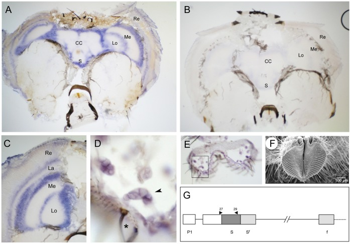
Figure 5. Md-fru P1 transcripts are expressed in the CNS and in peripheral sensory organs.
(A) Frontal section of a male head hybridised with Md-fru P1-specific antisense RNA probe. Strong ubiquitous staining is observed in the layers below the retina and around the optic lobes (Re: Retina, La: Lamina, Me: Medulla, Lo; Lobula), as wells as around the central complex (CC) and the subesophageal ganglion (S). (B) Frontal section of a male head hybridised with the Md-fru P1 sense RNA probe. (C) Higher magnification of the stained areas in the optic lobes. (D) Close-up of basal neurons (arrowhead) connected to a sensory bristle (black star) located in the labellum and expressing Md-fru P1 transcripts. (E) Overview of the sectioned labellum shown in D (boxed). (F) SEM imaging of the fly’s mouthpart (labellum) and surrounding sensory bristles. (G). Primers used to prepare templates for P1-specific sense and antisense RNA probes are indicated as black triangles (Md-fru-27 and Md-fru-29).

