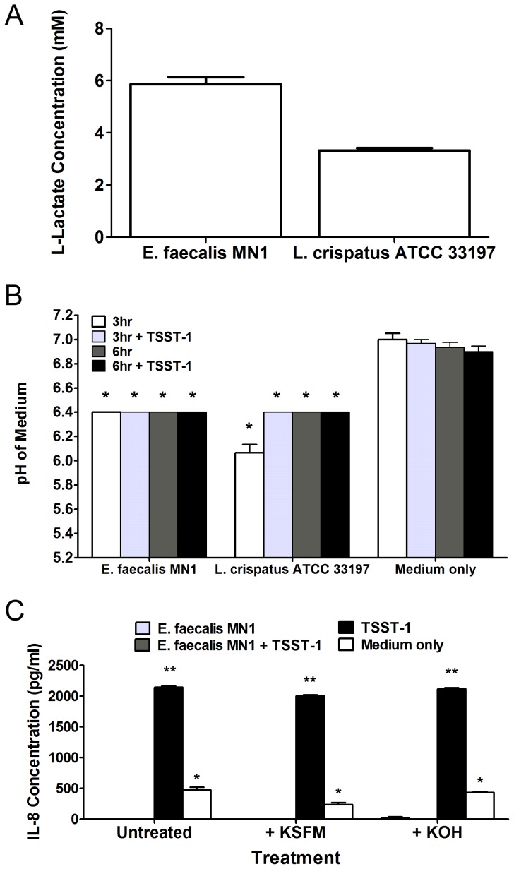
Figure 5. Lactic acid is not solely responsible for IL-8 inhibition.
A) E. faecalis MN1 and L. crispatus ATCC 33197 (both at 1×10⁷ CFU/well) were incubated with TSST-1 (100 µg/ml) on HVECs for 6 h, and lactic acid was measured in the tissue culture medium using a colorimetric assay. B) E. faecalis MN1 and L. crispatus ATCC 33197 (both at 1×10⁷ CFU) were incubated ± TSST-1 (100 µg/ml) with HVECs for 6 h, and the pH of the tissue culture media was measured after 3 and 6 h. All conditions with enterococci or lactobacilli were significantly lower than the TSST-1 or medium only controls at 3 h and 6 h by individual Student's t tests (p<0.001). C) Cells were incubated with E. faecalis MN1 (1×10⁷ CFU) ± TSST-1 (100 µg/ml) for 6 h. After 3 h, cells were either left untreated or were treated with additional KSFM tissue culture medium or 1M KOH to neutralize the pH of the medium. IL-8 was measured after 6 h, at which point the pH of the medium in treated samples was still at or near neutral. All conditions that contained E. faecalis MN1, no matter the pH level, showed significantly lower levels of IL-8 than that of TSST-1 alone. **Significantly higher than all other conditions and *significantly higher than conditions containing E. faecalis MN1 irrespective of neutralization treatment by one-way ANOVA [F(3,8) = 555.1, p<0.0001] and Tukey's post-hoc test. N = 3 replicates for each experiment.

