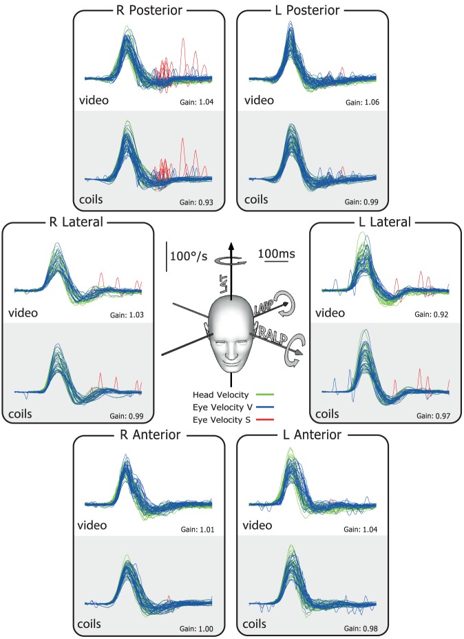
Figure 2. Simultaneous video and search coil head impulse recordings of all semicircular canals in a healthy subject.
The six double panels in each figure show simultaneous measures by scleral search coils (lower half of each panel) and the video head impulse system (upper half of each panel). The records here show that for a healthy subject the traces of head and eye velocity are almost superimposed for the direction of each semicircular canal. The VOR gain values for head turns in every canal plane are in the normal range. The plots themselves in this and the following figures are time series showing superimposed records of the head velocity stimulus (head velocity – green traces) and the slow-phase eye-velocity responses (eye velocity VOR – blue traces) to about 20 brief unpredictable head turns in the direction of each semicircular canal. Overt or covert saccades are shown as red traces [10]. Tiny overt catch-up saccades are normal in healthy subjects. In these figures eye velocity has been inverted to allow easy comparison with head velocity, and for purposes of illustration both leftward and rightward head movements are shown as positive. The average VOR gain value is shown next to each group of responses. The inset at the centre shows the rotation axes of the semicircular canals being tested.

