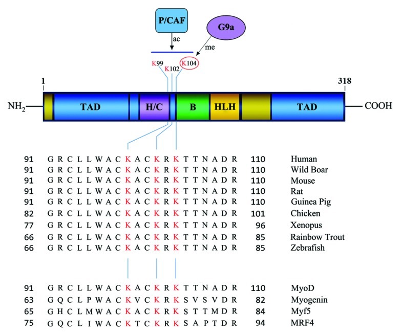
Figure 1. Schematic representation of the MyoD domain structure (upper panel). The basic (B) DNA-binding domain; helix-loop-helix (HLH) dimerization domain; transactivation domain(s) (TAD); and the cysteine-histidine rich region (H/C) are shown. Numbers indicate amino acid residues. Alignment of MyoD cDNA from various species (middle panel) show three highly conserved lysine (K) residues (highlighted in red) that are acetylated by P/CAF upon differentiation. K104 is methylated by G9a in undifferentiated myoblasts (numbering based on the human/mouse cDNA). These lysine residues are conserved in all MRFs (lower panel).
