
Fig. 2.
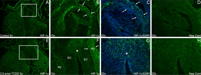
HIF-1α staining of control verses TCDD-treated embryos. Specific sites of immunofluorescent staining for HIF-1α near the OFT myocardium of a stage 30 chicken embryonic heart. HIF-1α nuclear-localized immunostaining intensity and frequency was high in the control (a–c) OFT myocardium (arrows in b, c) while lower in the TCDD-treated embryos (f–h). However, TCDD-treated embryos had increased HIF-1α staining within the endocardial cushions (g between arrows, h). My myocardium and EC endocardial cushions. Bright staining in d and h is autofluorescence of red blood cells and not that of nuclear-localized HIF-1α staining