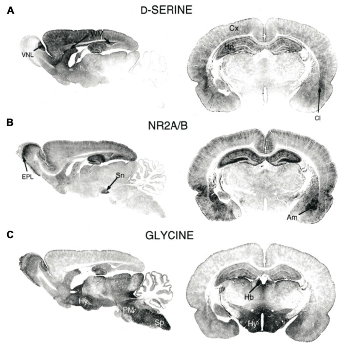
FIGURE 1.
Immunohistochemical staining of (A) D-serine, (B) NMDAR subunits GluN2A/B, and (C) glycine in rat brains (P21). Am, amygdala; Cl, claustrum; Cx, cortex; EPL, external plexiform layer; Hb, habenula; Hy, hippocampus; PM, pons/medulla; Sn, substantia nigra; Sp, spinal cord; WM, white matter; VNL, vomeronasal nerve layer. Figure modified from Schell et al. (1997).

