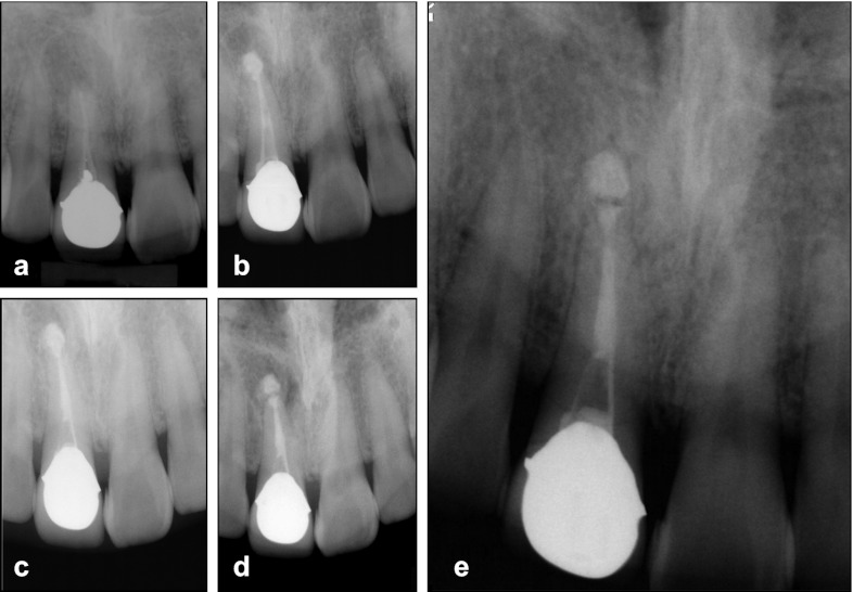
Figure 2.
Radiographs of case 2. (a) The first visit for diagnosis. (b) The MTA extrusion into the periapical lesion during the one-step apexification. (c) Completion of MTA placement. (d) 3-month recall showing the gradual healing of the apical lesion with radiolucent halo around the extruded MTA. (e) 54-month follow-up showing complete osseous repair with continuum of lamina dura-like structure along the extruded MTA. MTA, mineral trioxide aggregate.

