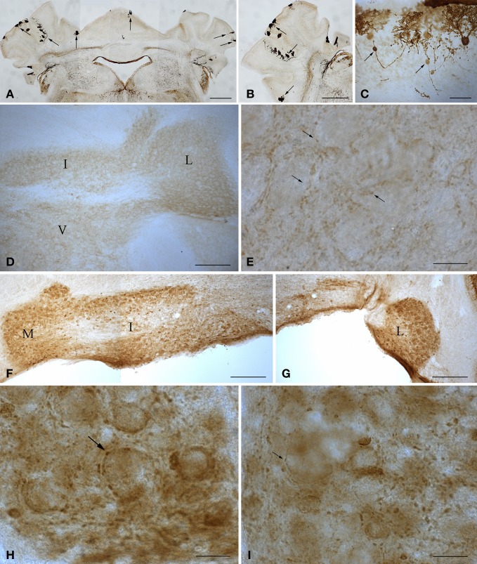
Figure 2.
Microphotographs of coronal (A, D–I) and sagittal (B,C) sections immunostained with anti-calbindin (A–C) and anti-parvalbumin (D–I) antibodies illustrate the main features of the cerebellum of wild type and 1-year-old tambaleante mice. Scarce Purkinje cells remain throughout the cortex (A–B, arrows), preserving a mirror location on both sides of the cerebellar cortex (A, arrows). Axons of degenerating Purkinje cells show typical axonal torpedoes (C, arrows). Parvalbumin immunoreactivity is restricted to small endings (E, arrows), and is absent in both deep cerebellar and vestibular nuclei neuronal somata (D), in wild-type cerebellum. In tambaleante cerebellum, parvalbumin immunoreactivity is present in neuronal somata of deep cerebellar nuclei (F,G), and in large (H, arrow) and small (I, arrow) terminal endings. I, L, and M, interposed, lateral, and medial cerebellar nuclei. V, vestibular nuclei. Bar = 500 μm (A,B), 200 μm (D,F,G), 30 μm (C), and 20 μm (E,H,I).

