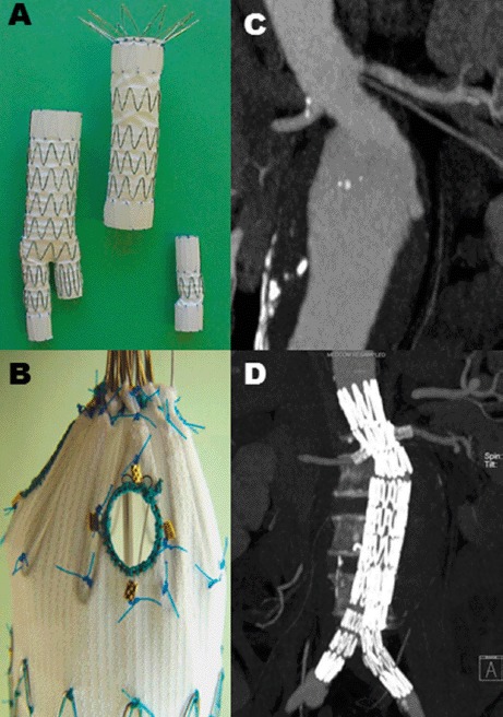
Fig 7.

An in vitro deployed fenestrated stent-graft (Image A). Image B – a magnified view demonstrating a pre-planned fenestration within the proximal component. Image C – a preoperative CT scan of patient with a short and angulated aortic neck that is unsuitable for standard EVAR. Image D - postoperative CT scan (same patient as Image C) showing a deployed fenestrated stent-graft with exclusion of the AAA and patent visceral arteries
