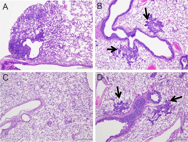
Fig 3.
Histological analysis of lung tissues. Three mice per group were intranasally infected with 1,000 TCID50 (>10 MLD50) of A/Osaka/129/2009 (A/H1N1pdm). The mice were treated from 1 h p.i. for 15 days and were autopsied on day 15 p.i. to collect lung tissues. Representative pictures with H&E staining of each group are shown. (A) Normal infected mice; (B) immunosuppressed and infected mice treated with 0.5% methylcellulose (control); (C) immunosuppressed and infected mice treated with peramivir (40 mg/kg/day); (D) immunosuppressed and infected mice treated with oral oseltamivir (10 mg/kg/day). Arrows indicate metaplasia.

