Fig 1.
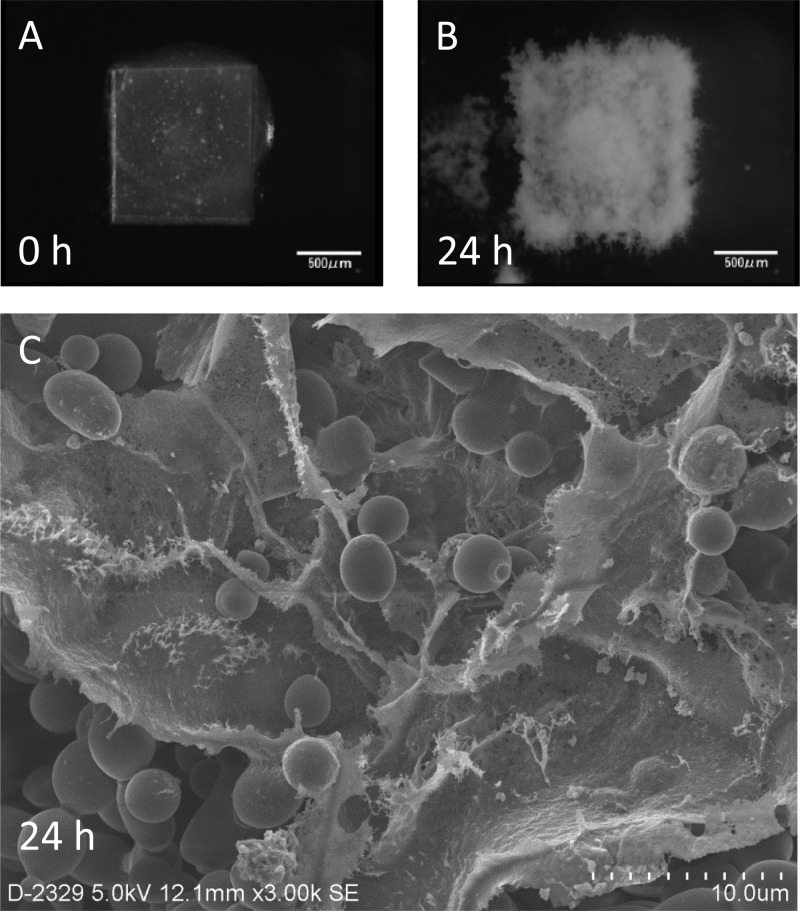
Development of Candida albicans biofilms on a silicon disk in flow, observed from the top, and the electron microscopic image. (A and B) Attachment phase (A) and 24-h-old mature biofilms (B). (C) Mature biofilms were also observed under a scanning electron microscope, revealing a dense extracellular matrix covering the accumulated Candida cells.

