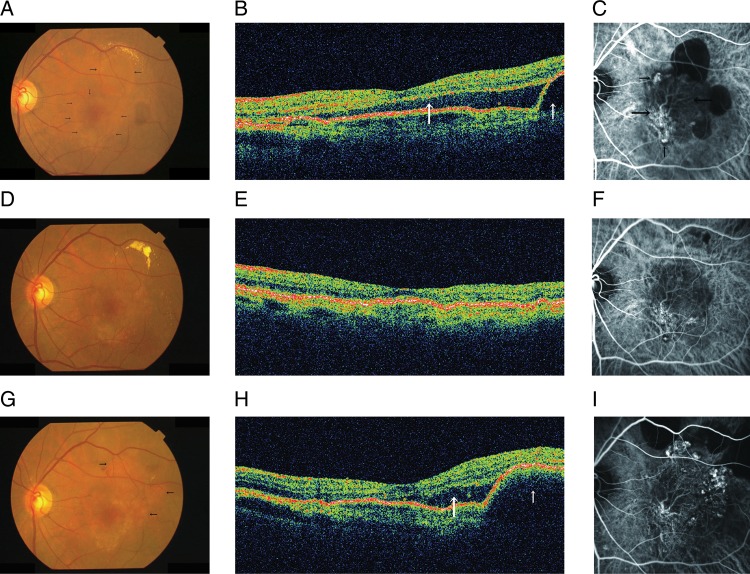
Figure 3.
The left eye of a 71-year-old man with polypoidal choroidal vasculopathy. (A) At baseline, a fundus photograph shows a serous retinal detachment (SRD) involving the fovea (small arrows) and a serous pigment epithelial detachment (PED) (large arrows). (B) A horizontal optical coherence tomography (OCT) image shows a subfoveal SRD (large arrow) and PED temporal to the fovea (small arrow). (C) indocyanine green angiography (ICGA) shows polypoidal lesions (small arrows) and a branching vascular network (BVN) (large arrows). The planimetric size of the BVN is 4.33 mm2. (D and E) One year after the first injection of ranibizumab, a fundus photograph and OCT shows resolution of the SRD and PED. (F) ICGA shows regression of polypoidal lesions and no increase in the size of the BVN (4.42 mm2). (G) Two years after the first injection, a fundus photograph shows some elevated orange-red lesions (arrows). (H) A horizontal OCT image shows a recurrent subfoveal SRD (large arrow) and a PED temporal to the fovea (small arrow). (I) ICGA shows progression of the polypoidal lesions and the size of the BVN (5.75 mm2).

