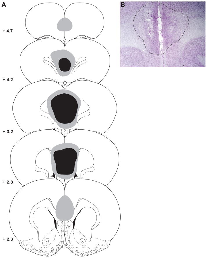
Figure 1.
The location and extent of ibotenic acid lesions of the medial prefrontal cortex (PFC). A) Schematic showing the largest (grey) and smallest (black) extent of the lesion on any particular section through the medial PFC. B) Photomicrograph depicting the average extent of damage caused by ibotenic acid infusions. Adapted from [24].

