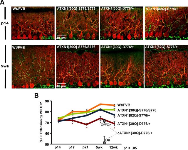
Figure 1.
Altered CF development in SCA1-affected mice. A, Immunofluorescent confocal images taken at 60× from p14 (top) and 5 weeks of age (bottom) cerebellar sections immunostained with calbindin to identify PCs (red) and VGLUT2 to identify CF terminals (green). B, Time course of distal extension profile of CF terminals onto the PC dendrites at different stages in development. The Wt FVB and ATXN1[30Q]-S776/S776 mice have a steady progression until 5 weeks of age. SCA1 mice show no progression (ATXN1[30Q]-D776/+) or retarded progression (ATXN1[82Q]-S776/+). Number of animals (n) assessed for each genotype are Wt FVB: p14, 4; p17, 4; p21, 4; 5 weeks, 3; 12 weeks, 5; ATXN1[30Q]-S776/S776: p14, 4; p17, 4; p21, 3; 5 weeks, 3; 12 weeks, 7; ATXN1[82Q]-S776/+: p14, 3; p17, 3; p21, 3; 5 weeks, 3; 12 weeks, 6; and ATXN1[30Q]-D776/+: p14, 3; p17, 4; p21, 4; 5 weeks, 3; 12 weeks, 4. At 7 weeks cATXN1[30Q]-D776 gene-on, n = 3; and cATXN1[30Q]-D776 gene-off/gene-on, n = 4. Data are represented as mean ± SEM. *, denotes a significant difference compared with the Wt FVB controls (p < 0.01, Bonferroni post hoc comparison). The 12 weeks of age time point data are provided as a point of reference from Duvick et al., 2010.

