
Figure 5.
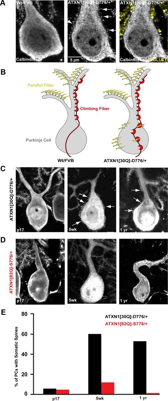
Abnormal parallel fiber distribution in ATXN1[30Q]-D776 mice. A, 350× immunofluorescent images of calbindin-labeled PCs somata from Wt FVB, left and ATXN1[30Q]-D776/+ mice, middle and right. Atypical spines can be seen protruding from the PC soma and apical dendrite in ATXN1[30Q]-D776/+ (white arrows, middle). Colocalization between the presynaptic parallel fiber–terminal marker terminal marker, VGLUT1 (yellow, right), and somatic PC spines. B, Diagram depiction of the disruption of CF (red) and parallel fiber synapses (yellow) and their relationship to PC spines in Wt FVB and ATXN1[30Q]-D776/+ mice. C, D, 150× immunofluorescent images of spine development in SCA1 mice. ATXN1[30Q]-D776/+ mice (C) display somatic spines at 5 weeks of age and remain out to 1 year of age. D, No atypical spines were detected at 5 weeks of age in ATXN1[82Q]-S776/+ mice. E, Time course of the percentage of PCs displaying somatic spines. ATXN1[30Q]-D776/+: n = 50 PCs examined; ATXN1[82Q]-S776/+: n = 63.