
Figure 1.
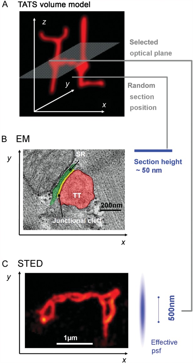
The cardiac cell membrane exerts deep intracellular functions through TT invaginations, forming a continuous three-dimensional TATS network composed of transversal and longitudinal components. (A) Minimal three-dimensional TATS volume model composed of 2 transversal TTs continuously connected with longitudinal TT elements; example imaging/sectioning planes in (B) and C) are indicated. (B) Individual TT–jSR contacts were investigated in control and HF samples in end-stage DCM/ICM by Zhang et al.8 Increased spatial separation between Z-lines and jSR junctions (green) was shown among other observations in human HF samples. While ultrathin sectioning allows for precise EM measurements of TT–jSR membrane architectures, sectioning occurs randomly relative to the complex three-dimensional membrane structures. (C) STED image of deep intracellular TT membrane structures specifically labelled with di-8-ANEPPS showing abnormally enlarged TT cross-sections and TATS network remodelling 4 weeks post-MI in mouse hearts.4 Optical planes can be directed to specific objects (e.g. transverse-axial TATS intersections) in the focal plane based on image contrast. Image analysis based on identified objects can be semi-automated for quantification. The effective psf indicates a lateral resolution of ∼55 nm and a z resolution of ∼500 nm, the latter limiting 3D resolution similar to confocal imaging here; imaging by z stacks allows for targeted three-dimensional sampling. The Field of view can be adapted to the TATS network dimensions or individual TT objects as needed. B and C reproduced with permission.