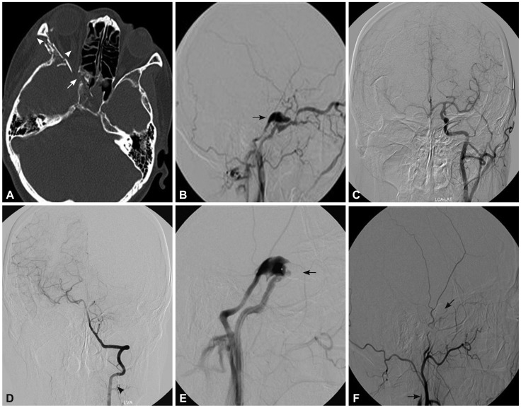
Fig. 3.
A 42-year-old woman who presented with right-sided proptosis, chemosis, orbital bruit, and headache 35 days after a vehicle accident. A: Computed tomography indicated right-sided proptosis, superior ophthalmic vein enlargement (arrowhead), and multiple orbital fractures (arrow). B: Cerebral angiography confirmed a high-flow direct CCF that drained into the superior ophthalmic vein and inferior petrosal sinus with cortical reflux (arrow). C and D: Cerebral angiography of the contralateral ICA confirmed good compensation of the circle of Willis. E: Cerebral angiography of the ipsilateral ICA after two-balloon embolization confirmed partial occlusion of the fistula and absence of the distal ICA (arrow), which prompted a diagnosis of obliteration of the ipsilateral ICA distal to the fistula point. F: The CCF was successfully treated after subsequent occlusion of the proximal ICA with a detachable balloon (arrow). CCF: carotid-cavernous fistula, ICA: internal carotid artery.

