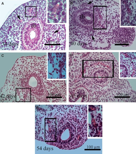
Fig. 2.

Histological sections of human lung stained with H/E. The insets correspond to higher magnification of the area enclosed in the rectangles. (A,B,C) The capillary plexus (arrows) around a lung terminal bud (tb) of embryos aged 34, 40 and 42 days pf. Primitive erythroblasts are seen within the lumen of some blood vessels (inset). (D) Primitive erythroblasts arranged in a single line within the capillary network of the lung (inset) at day 45 pf. From this embryonic age on, an increase of the amount of blood cells in the distal vascular plexus as well of their diameter was observed. Note that some definitive erythrocytes can be seen within the capillaries (inset) (E).
