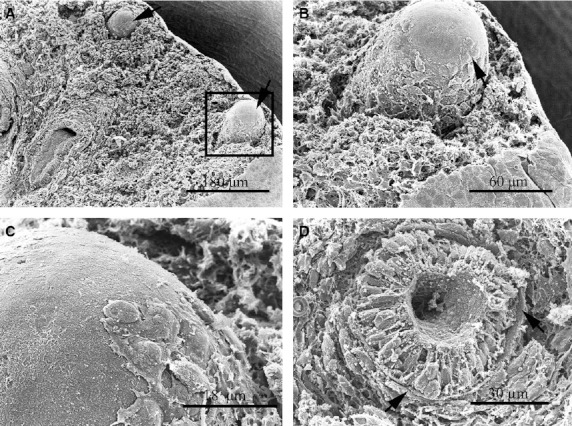
Fig. 3.

SEM images of the human lung at day 50 pf. (A) Two lung buds at the periphery of the lung (arrows). (B) Higher magnification of the tip of the blind projection of the lung bud enclosed in (A). The tip of the lung bud appears covered by a basal membrane and no mesenchymal cells were observed at this location. A higher magnification of the tip image is shown in (C). Some mesenchymal cells that look like migratory cells are attached to the basal lamina. (D) A transverse section through a terminal bud, showing a very regular-shaped endodermal epithelium surrounded by a strong basal lamina (arrows). Note that the mesenchymal tissue forms a sheath around the bud.
