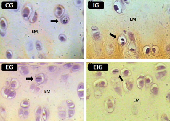
Fig. 1.

Histological sections of the femoral articular cartilage from each of the four groups of rats (C, I, E, and EI) were used to measure the major and minor nuclear diameters of chondrocytes (arrows). EM, ECM. Figures are from the intermediate zone of the cartilage. HE staining. Scale bar: 30 μm.
