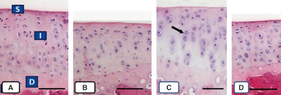
Fig. 3.

Microphotographs of sections of cartilage sectioned perpendicular to the articular surface from rats in groups C (A), I (B), E (C), and EI (D). Observations of the superficial (S), intermediate (I), and deep zones (D) were clearly differentiated in (A), (C), and (D), but not in (B). Two chondrocytes are indicated in (C) with arrows. Scale bars: 100 μm.
