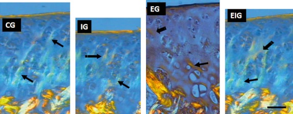
Fig. 4.

Light-field photomicrographs via polarization microscopy of the femoral cartilage sections from groups C, I, E, and EI stained with Picrosirius to observe the changes in collagen (arrows) in the immobilization (I) and exercise (E and EI) groups. Collagen appears as orange, white, and green vertical fiber bundles on a blue background above the cancellous bone. Collagen concentration seemed to have been reduced in group I and augmented in group E in relation to the other groups. Scale bar: 30 μm.
